Detection limits of automated MRI morphometry for phenotyping in the rodent brains for applications in neurological disorders
Date:
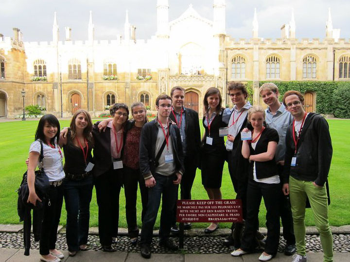
I presented a poster at the Amgen Scholars European Symposium 2010 at the University of Cambridge, describing research conducted during my summer internship at the Wolfson Brain Imaging Centre under the supervision of Adrian Carpenter and Steve Sawiak. The symposium brought together undergraduate researchers from institutions across Europe to share summer projects and participate in a series of academic talks and poster sessions.
This presentation was part of the broader Amgen Scholars Programme, a global undergraduate research initiative launched by Amgen. As reported at the time, the programme had a 6% acceptance rate in 2010.
My project focused on evaluating the detection limits of automated voxel-based morphometry (VBM) for identifying structural changes in rodent brains using high-resolution magnetic resonance imaging (MRI). Working with simulated 3D phantom images and controlled morphometric changes, we studied how factors such as smoothing, effect size, spatial extent, and sample size influence the sensitivity and specificity of VBM-based statistical tests.
The poster detailed our framework for generating synthetic datasets, preprocessing and segmentation of MRI, and performing voxel-wise statistical inference under various noise conditions and anatomical configurations. This work served as a methodological contribution to understanding how experimental design choices affect the detectability of subtle neuroanatomical changes in small-animal imaging studies, informing both phenotyping pipelines and the interpretation of rodent MRI experiments.