Advanced MRI Techniques for Early Detection of Brain Metastases in Small Cell Lung Cancer
Date:
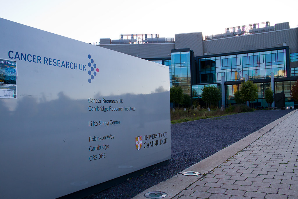
I delivered an oral presentation at the Cancer Research UK Cambridge Research Institute (CRI) summarizing the results of my summer research internship in the laboratory of Professor John Griffiths at the University of Cambridge. The project focused on evaluating advanced magnetic resonance imaging methods for the early detection of brain metastases in small cell lung cancer (SCLC).
Working under the supervision of Professor Griffiths and Dr Dominick McIntyre, I developed image analysis methods for a mouse model of SCLC brain metastasis. The aim was to determine whether subtle changes in normal appearing brain tissue could be detected with quantitative MRI before metastatic lesions become visible on conventional contrast enhanced scans. This involved processing serial brain images, extracting magnetization transfer, diffusion and spectroscopy measurements, and registering all scans to the high resolution SPMMouse atlas to allow consistent examination of predefined brain regions across time.
In the presentation, I described early findings showing how quantitative imaging parameters evolve in regions that later develop metastases and how these regions compare with contralateral tissue that appears normal on visual inspection. The work demonstrated the potential of advanced MRI methods to identify very early metastatic changes, contributing to ongoing efforts in the Griffiths lab to develop non invasive imaging biomarkers that could guide individualized treatment decisions for patients with SCLC.
You can see some photos taken during my internship at CRUK Internship (flickr).